 When used appropriately imaging can provide valuable information about the different structures within our bodies. Unfortunately, imaging is only one piece of the puzzle and to form the whole picture first we need to think about the injury history and have a physical assessment. It is important to remember that imaging alone does not give a diagnosis and should not be relied upon. Many people with no pain or dysfunction will have abnormal findings on imaging. Read more here to help you decide on whether or not you need a scan.
There are many different types of scans that are used to look at musculoskeletal structures. In part one of this blog I am just going to discuss x-ray and CT scans. Part two will be MRI and ultrasound scans.
X-ray
This is a form of electromagnetic radiation. These high energy x-rays can pass through objects such as the body.
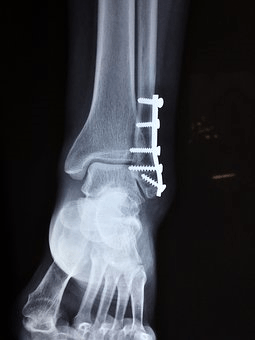
When an x-ray is performed, the body is positioned between the x-ray source and an x-ray detector. When turned on x-rays travel through the body and each tissue type absorbs different amounts depending on its radiological density. This is determined by density and the atomic number (how many protons are in the atom’s nucleus) of the tissue. Bone for example has a higher radiological density than fat or muscle and it absorbs x-rays more easily. Therefore, bone shows up white against a black background.
What is it used for?
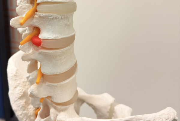
X-rays are commonly used to look at bony alignment, abnormalities or fractures. They can also be used to look for calcifications, some tumours or masses and the presence of foreign objects.
What are the risks?
Generally, when used well the benefits of an x-ray should out way the risks. In saying that x-rays produce ionising radiation that can harm tissues. The risk of damage from this radiation increases with the number of x-rays you have had. Children are generally more sensitive to this radiation. In pregnant women, x-rays of the abdomen and pelvis are generally avoided to prevent any increased risk for the unborn child.
CT scan (Computed Tomography)
A CT scan is basically a computerised x-ray. Unlike a normal x-ray it uses a motorised x-ray source that can move. This rotates around a circular donut shaped opening which the body is passed through. Cross sectional images of the body are generated that are more detailed than a normal x-ray. As the CT scanner takes many cross-sectional images or slices, they can be displayed individually, or they can be digitally put together to form a three-dimensional image.
What is it used for?
A CT scan is used for imaging complex bony fractures or tumours in much more detail than normal x-ray. It can also be used to screen for possible tumours, various types of heart disease, haemorrhage, some blood clots and many other conditions.
What are the risks?
Like x-rays, CT scans use ionising radiation therefore it carries the same type of risks.
Click here for Imaging Explained Part Two: All about MRI and Ultrasound scans.